
Figure 5. Immunoblots of EGFR family members in 2D and 3D cultures.
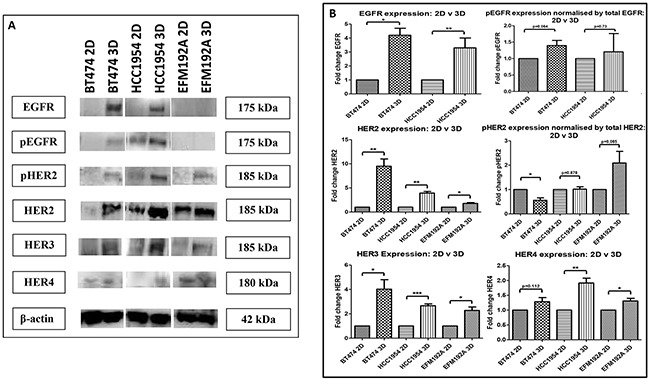
A. EGFR, pEGFR, HER2, pHER2, HER3 and HER4 were all increased in 3D cultured cells compared to their 2D counterparts, with the exception that EGFR/pEGFR were undetected in EFM192A cells grown in either format. B. Densitometry of respective immunoblots. Graphs represent triplicate biological repeats and are displayed as mean ± SEM, where *p < 0.05; **p < 0.01; **p < 0.001.