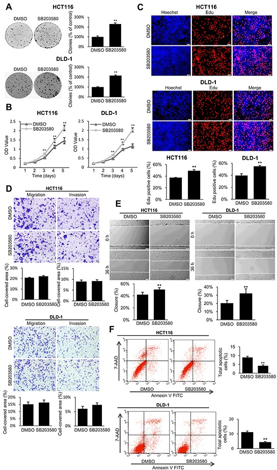
Figure 7. The p38 inhibitor reversed the tumor suppressive effect of TES in CRC cells.

A. Representative photographs of cell culture plates following staining with crystal violet for colony formation of the TES-overexpressing HCT116 and DLD-1 cells in the presence or absence of p38 inhibitor. Number of colonies was quantified. B. The cell growth of the TES-overexpressing HCT116 and DLD-1 cells in the presence or absence of p38 inhibitor were determined by MTT assays at each time point. C. Representative profiles of EdU cell proliferation assay of the TES-overexpression HCT116 and DLD-1 cells in the presence or absence of p38 inhibitor. Representative photographs were taken at 200× magnification. D. Cell migration and invasion was assessed after 24 h incubation in the presence or absence of p38 inhibitor by transwell assays. Representative photographs were taken at 100× magnification. E. Representative images of wound healing assay by scraping culture dishes using a pipet tip and closure in the presence or absence of p38 inhibitor after 36 h of culture. Representative photographs were taken at 100× magnification. F. Cell apoptosis of TES-overexpressing HCT116 and DLD-1 cells in the presence or absence of p38 inhibitor was measured by flow cytometric analyses. Representative biparametric histogram showing cell population in apoptotic (top right and bottom right quadrants), viable (bottom left quadrant) and necrotic (top left quadrant) states. ** p < 0.01. Data are plotted as the mean ± SD from five independent experiments. Bars indicate the standard deviation of the mean.
