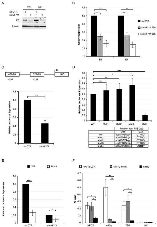
Figure 4. NF-Y transcriptionally controls the expression of HPV18-URR driven genes.

A. Western Blot analysis of E6 protein in whole cell extracts from Hela infected with shCTR and shNF-YA for 72h and 96h. Tubulin was used as loading control. B. Relative expression levels of E6/E7 genes normalized to the hRpl19 transcript in shNF-YA cells versus shCTR, arbitrarily set at 1. Statistical significance was determined with independent t-test (** p < 0.01). C. Upper panel: schematic representation of CCAAT boxes position in HPV18-URR, cloned upstream of the luciferase (LUC) reporter gene. Lower panel: relative HPV18-URR-driven luciferase activity in shCTR and shNF-YA cells. Statistical significance was calculated with independent t-test (** p < 0.01). D. Relative luciferase expression of mutant promoters with respect to wt HPV18 promoter. Statistical significance was calculated with independent t-test (* p < 0.05; **** p < 0.0001). The table indicates the position and sequence of the two wt and mutated NF-Y-motives. E. Relative luciferase activity of wt and mut4 HPV18 URR in shCTR and shNF-YA cells. F. ChIP analysis of NF-YA, c-FOS and TBP binding to HPV18-LCR, c-Myc promoter and negative control region (CTRL-) in Hela cells. Enrichment was calculated as percentage of IP recovery. Statistical significance was calculated with independent t-test between promoters of interest and CTRL- region (* p < 0.05; ** p < 0.01). Error bars indicate SEM.
