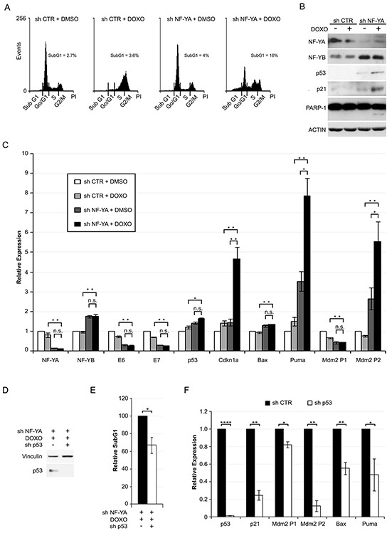
Figure 6. NF-YA loss sensitizes Hela cells to Doxorubicin-induced p53-dependent cell death.

A. DNA distribution analysis of Propidium Iodide-stained Hela cells infected with shCTR and shNF-YA for 72h and then treated with DMSO or 0.1 μM Doxorubicin (DOXO). The percentages of SubG1 events are indicated. Shown images are representative of three independent experiments. B. Western blot analysis of whole cell extracts in the experimental conditions described above. Antibodies are indicated. Actin was used as loading control. C. qRT-PCR relative expression analysis of p53 target genes in shCTR and shNF-YA cells treated or not with DOXO. The housekeeping hRpl19 gene has been used for normalization. The expression levels of control cells (shCTR +DMSO) have been arbitrarily set at 1. D. p53 expression levels in NF-YA-inactivated cells infected with shCTR and shp53 and treated with DOXO. E. Effects of p53 loss (shp53) on SubG1 events in NF-YA-inactivated cells treated with DOXO. The percentage of SubG1 in NF-YA-inactivated cells infected with shCTR has been arbitrarily set at 100%. F. qRT-PCR analysis of the indicated transcripts in NF-YA/p53 double knocked down cells versus NF-YA-inactivated cells (set at 1), following DOXO treatment. Statistical significance was calculated with independent t-test (* p < 0.05; ** p < 0.01; **** p < 0.0001;). Error bars indicate SEM.
