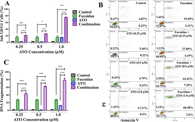
Figure 2. Apoptosis assays.

NB4 cells were treated with increasing doses of ATO with or without fucoidan (20 μg/ml) and various assays conducted after 48 hours. A. DNA content was analyzed and sub G0/G1 population representing dead cells was calculated. Mean ± SEM values of three replicates are shown. Statistical significance was determined by two-way ANOVA, followed by Bonferroni post-test. B. Representative annexin V/PI apoptosis assay. The amount of apoptotic cells (positive Annexin V) increased when fucoidan was combined with ATO at both low and high doses. Mean ± SEM values of three replicates are shown. C. DNA fragmentation. The amount of apoptotic cells with fragmented DNA significantly increased when fucoidan was combined with ATO at both low and high doses. Mean ± SEM values of three replicates are shown. Statistical significance was determined by two-way ANOVA, followed by Bonferroni post-test (***: p<0.001, **: p<0.01, *: p< 0.05).
