
Figure 7. MA-mediated ER stress increased cell death in astrocytes.
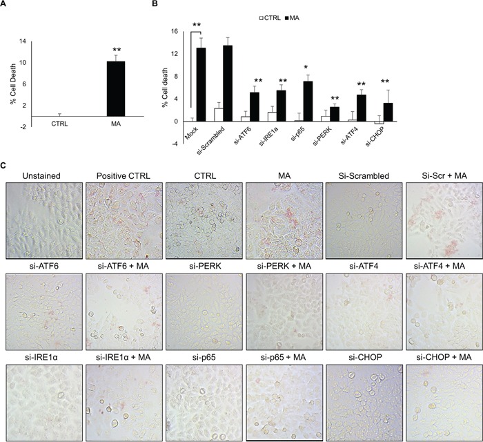
Cell death was measured at 48 hours after the treatment with 500 μM MA. Briefly, SVGA cells were seeded at 6 × 10⁵ cells in a 6-well plate and transfected with various siRNA as mentioned in methods. After 48 hours, the cells were reseeded in 12-well plate and allowed to adhere overnight. Finally, these cells were treated with 500 μM MA for additional 48 H and MTT assay was performed to assess the effect of ATF6, PERK, ATF4, IRE1α, p65 and CHOP knockdown. A. The % cell death was calculated considering the absorbance in untreated control as 100% viability. B. The involvement of ATF6, IRE1α and PERK pathways and their respective intermediates leading to CHOP in cell death was assessed using MTT assay after knockdown of each of the intermediates with siRNA, which demonstrated reduction in MA-mediated cell death at variable extents. C. Similarly, to confirm these results, TUNEL staining was performed at 48 hours after MA treatment. The TUNEL staining was performed using TACS-XL In Situ Apoptosis Detection Kit - DAB kit as described in materials and methods. The images were captured at 20X zoom using Labomed iVu5100 camera. The results shown in bar graphs were obtained from at least 3 independent experiments with each treatment performed in triplicates and presented in mean ± S.E. Statistical significance was calculated using one-way ANOVA with multiple comparisons and the values were considered significant if p-value ≤ 0.05 (*) or ≤ 0.01 (**).