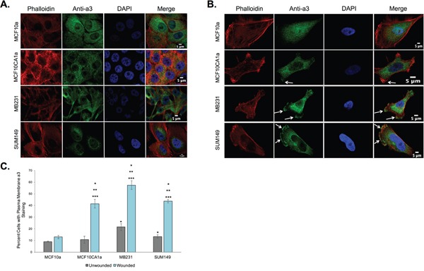
Figure 1. Subunit a3 localizes to the plasma membrane of human breast cancer cells.

MCF10a, MCF10CA1a, MB231, and SUM149 cells were grown to confluence on poly-d-lysine coated coverslips. To assess the effects of cell migration on a3 localization, a wound was made in the cell monolayer and cells were allowed to migrate for 4 h. Cells were then fixed, permeabilized, and immunostained using antibodies against subunit a3 of the V-ATPase and Alexa Fluor® 568 phalloidin to stain actin followed by incubation with secondary antibodies as described under Materials and Methods. Images were taken with identical exposure times and antibody concentrations. A. Shown are representative images of confluent MCF10a, MCF10CA1a, MB231, and SUM149 cells stained for phalloidin (left), subunit a3 (second from left), DAPI (second from right), and the merged images (right). B. Shown are representative images of MCF10a, MCF10CA1a, MB231, and SUM149 cells (with monolayer wounded) stained for phalloidin (left), subunit a3 (second from left), DAPI (second from right), and the merged images (right). White arrows indicate plasma membrane subunit a3. C. Quantification of plasma membrane staining for subunit a3 in confluent or migrating cells. To quantitate plasma membrane staining, 50-80 cells from three separate experiments were counted and the number of cells showing plasma membrane a3 localization was determined. The graphed data represents the percentage of cells showing a3 localization to the plasma membrane. All error bars indicate S.E. *, p < 0.05 relative to unwounded MCF10a cells. **, p < 0.05 relative to wounded MCF10a cells. ***, p < 0.05 relative to the unwounded cell of the same cell type.
