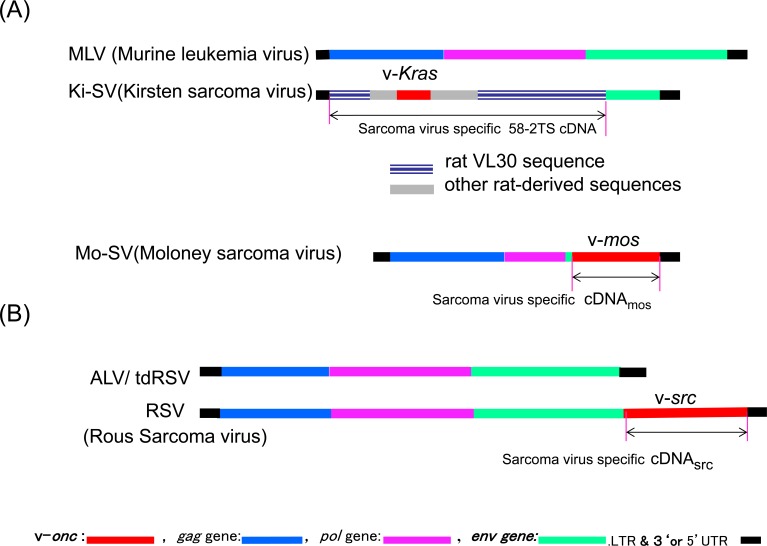
Figure 2. Comparisons of Ki-SV, Mo-SV, RSV, and the corresponding leukemia viral genomes.
A. Genomic structures of Murine leukemia virus (top structure), Kirsten sarcoma virus [17, 72], and Moloney sarcoma virus [141]. B. ALV/tdRSV (avian leukemia virus/transformation defective RSV, the upper structure) and RSV (bottom structure). Leukemia viruses/tdRSV mutant genomes are shown for comparison. Each sarcoma virus figure shows regions covered by sarcoma virus-specific cDNA by an arrow.

