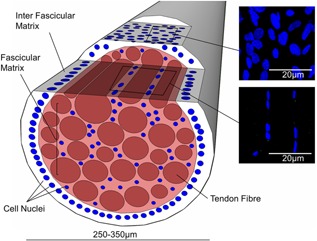
Figure 1.

Schematic showing a fascicle; highlighting the imaging planes in which confocal images are taken of the fascicular and inter‐fascicular matrix. The images on the right show representative images of nuclei and cilia in the inter‐fascicular and fascicular matrix.
