Figure 1.

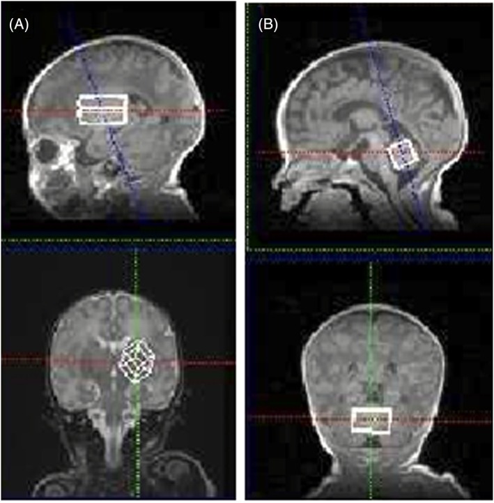
Example T 1‐weighted images (3D: T E/T R/T I 2.14/1570/800 ms) used for VOI selection of the BG (volume 4.4 mL) A, and cerebellum (3.7 mL) B, in a neonate (postconceptional age of 39 weeks)

Example T 1‐weighted images (3D: T E/T R/T I 2.14/1570/800 ms) used for VOI selection of the BG (volume 4.4 mL) A, and cerebellum (3.7 mL) B, in a neonate (postconceptional age of 39 weeks)