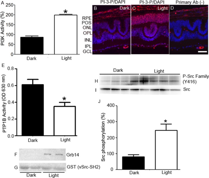
Figure 2. Light-dependent regulation of IR signaling proteins in cones.
Retinal lysates from dark- and light-adapted (300 lux, 30 min) Nrl−/− mice were immunoprecipitated with anti-IR antibody, and measured for PI3K activity using PI-4,5-P2 and [γ32P]ATP as substrates. The radioactive spots of PI-3,4,5-P3 were scraped from the TLC plate and counted A.. Data are means + SE (n = 4). *P < 0.001. The dark-adapted control (insulin receptor-associated PI3K activity) was set at 100%. Prefer-fixed sections of dark- B. and light-adapted C. Nrl−/− mouse retinas were stained for PI-3-P (red) and DAPI (blue). Panel D. represents the omission of PI-3-P antibody. RPE, retinal pigment epithelium; POS, photoreceptor outer segments; ONL, outer nuclear layer; OPL, outer plexiform layer; INL, inner nuclear layer; IPL, inner plexiform layer; GCL, ganglion cell layer. PTP1B activity was measured in the retinas harvested from the dark- and light-adapted Nrl−/− mice E.. Data are means +SE, n = 5, *P < 0.05. Dark- and light-adapted Nrl−/− mouse retinal lysates (duplicate) were incubated with GST-vSrc-SH2 fusion protein, followed by GST pull-down assay. The bound proteins were immunoblotted with anti-Grb14 F. and anti-GST G. antibodies. Retinas from four dark- and light-adapted Nrl−/− mice were immunoblotted with P-Src family (Y416) H. and Src I. antibodies. Quantified data (phosphorylated Src normalized to total Src) are means ± SE (n = 4), *p < 0.05 J.. The dark-adapted control was set at 100%.

