
Figure 4. Immunohistochemistry of allograft tumors.
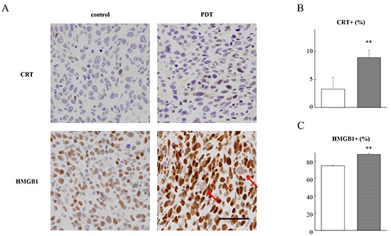
CT26 cells were inoculated into the dorsal skin of mice. Tumor-bearing mice were intravenously injected with 1.25 μmol/kg G-chlorin and, after 4 hours, were illuminated with 660-nm LED light (15 J/cm2). The tumors were excised and fixed in formalin for immunohistochemical examination at 3 hours after treatment with PDT plus G-chlorin. Representative immunohistochemical findings for lesions in the tumors of the control and PDT-treated mice (Panel A. original magnification ×400; scale bar = 50 μm). CRT B. and HMGB1 C. labeling indices in tumors. Data are the mean ± SD. Significance was determined by Welch's t-test. ** P < 0.01 relative to control.