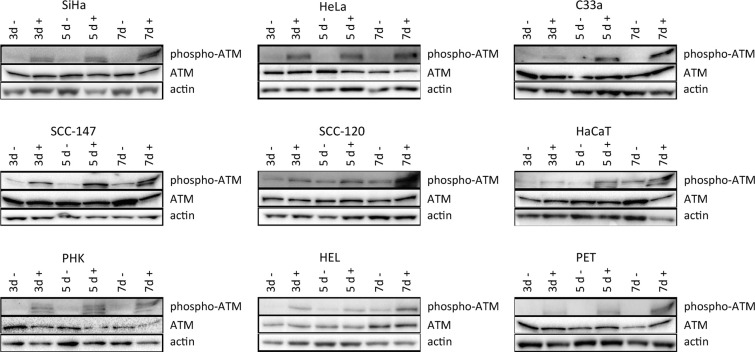
Figure 5. Western blot analysis of DNA damage signaling.
Protein extracts were obtained at 3, 5 and 7 days of no treatment or treatment with 50 μg/ml CDV. Afterwards, Western blotting was performed. Representative Western blots for each cell line after immunoblotting with ATM and phospho-ATM are shown. Actin was used as a loading control.

