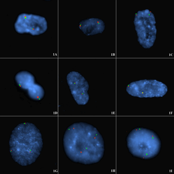
Figure 1.
A-I Intrachromosomal tethering of the subtelomeres of each single homologue in diploid and triploid non-cycling interphase nuclei at G1. FISH of diploid (A-F) or triploid interphase nuclei (G-I) from the following cell lines: CG04-0743BBRS (diploid) derived from skin and CG01-2042YA (triploid) derived from CVS. These non-cycling cells were probed with p-subtelomeric probe (labelled with spectrum orange) and q-subtelomeric probe (spectrum green) for (A) chromosome 4; (B) chromosome 5; (C) chromosome 7, (D) chromosome 10, (E) chromosome 17, (F) chromosome 20, (G) chromosome 18, (H) chromosome 12, (I) chromosome 6. The proportion of p-q tethered signals is shown in Table 2. In each case the majority of cells (76%–85%) showed pairing of short-arm and long-arm subtelomeres from single homologues often arranged on opposite sides of the interphase nucleus. Note also that an interphase topology is exhibited such that oval rosettes of chromatin can be seen in the present study in figs 1H, 1I, an elongated rosette in fig 1C, and off-centre rosettes in figs 1A, 1B.

