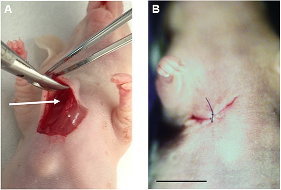
Figure 2. Establishment of Ewing's sarcoma PDOX model.

A. After making a skin incision on the right chest wall of a nude mouse, the space between the pectoral muscle and intercostal muscle (arrow) was expanded. A 4 mm3 fragment of the patient tumor was implanted orthotopically into the space. B. The pectoral muscle and the skin were closed with a 6-0 nylon suture. Scale bar: 10 mm.
