
Figure 3. LACE1 expressed in LACE1 KD background physically interacts with p53 and promotes its increased mitochondrial accumulation and nuclear reduction.
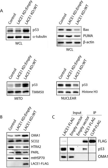
A. Overexpression of LACE1 leads to increased mitochondrial accumulation of p53 and its nuclear reduction. Whole-cell lysates (WCL), mitochondrial protein lysates (MITO) and nuclear extracts (NUCLEAR) were prepared from control cell line (Control) and from LACE1 KD cell line transfected with either the empty expression vector or LACE1-FLAG construct and subsequently immunoblotted with antibodies to p53, Bax and PUMA. Equal protein loading and fractionation efficiency was monitored using antibodies to TIMM50, histone H3 and α-tubulin. B. Loss of LACE1 leads to upregulation of PARL and downregulation of HTRA2. Mitochondrial protein lysates (MITO) were prepared from control cell line (Control) and from LACE1 KD cell line transfected with either the empty expression vector or LACE1-FLAG construct and subsequently immunoblotted with antibodies to OMA1, SCO2, HTRA2, FLAG, PARL and mtHSP70. C. P53 co-immunoprecipitates with LACE1-FLAG expressed in LACE1 KD background. LACE1 KD cells were transiently transfected with the empty expression vector, LACE1-FLAG construct or CLPP-FLAG construct. Mitochondrial fractions were solubilized with 1% Triton X-100 and subjected to anti-FLAG affinity purification. The bound antigens were eluted with 3x FLAG peptide solution under native conditions and processed for western blotting. Five percent (vol) of the purification input was loaded on the gels along with purified samples and immunoblotted with antibodies to FLAG, p53 and CLPP.