
Figure 4. Loss of LACE1 abrogates mitomycin c-induced translocation of p53 into mitochondria whereas its overexpression promotes mitochondrial translocation of p53.
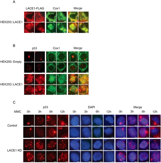
A-B. Overexpression of LACE1-FLAG in wild-type HEK293 cells promotes increased mitochondrial accumulation of p53. Cells grown on coverslips were transfected with the LACE1-FLAG construct or the empty vector and 48 hours post-transfection were fixed with 4% paraformaldehyde and permeabilized with 0.1% Triton X-100. Cells were then blocked by 10% Fetal Bovine Serum and primary detection was performed with antibodies to FLAG, p53 and COX1. After fluorescent secondary detection, cells were analyzed at 24°C using a Nikon Diaphot 200 inverted microscope equipped with a Plan-Apochromat 60×, numerical aperture 0.95, oil objective. The images were acquired with an Olympus DP50 CCD camera and Viewfinder Lite 1.0 software. Bar, 10 μM. C. Loss of LACE1 abrogates mitomycin c-induced mitochondrial translocation of p53. Cells grown on coverslips were treated with mitomycin c (MMC; 5g/mL) for 0, 3, 6 and 12h and then processed for immunofluorescence microscopy essentially as describe in (A-B) except that anti-p53 antibody and DAPI staining were used. Bar, 10 μM.