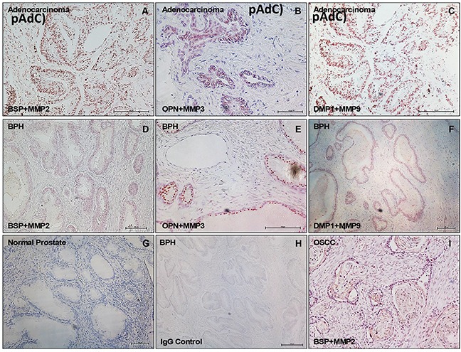
Figure 5. SIBLING-MMP interaction in prostatic neoplasms.

Top row panel is representative in situ proximity ligation assay (iPLA) showing SIBLING-cognate MMP in pAdC (Figures 5A-5C; red/brown dots), whereas middle row shows SIBLING-cognate MMP in BPH (Figures 5D-5F; red/brown dots). Third row show illustrative tissue negative control (normal prostate; Figure 5G), experimental negative control (BPH treated with pre-immune IgG; Figure 5H), and tissue positive control (human oral squamous cell carcinoma, OSCC; Figure 5I) known to co-express SIBLINGs and their cognate MMPs. Detection was with fluorescent/brightfield microscopy (20X). SIBLING-MMP interaction appears to be more intense in pAdC than in BPH. Other (non-cognate) SIBLING-MMP pairings were negative (see Supplementary Figure). Scale bar, 100 μm. Scale bar, 100μm.
