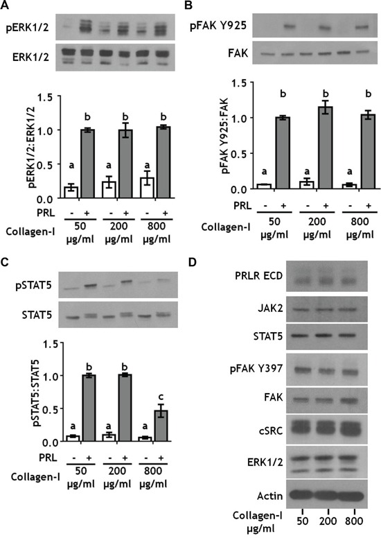
Figure 2. Collagen ligand density does not modulate PRL signals to ERK1/2 or FAK.

A-C. T47D were cells plated on 25 kPa polyacrylamide gels coated with either 50, 200, or 800 μg/ml collagen-I, serum starved for 24h, then treated ± PRL (4 nM) for 15 min. Cell lysates were immunoblotted with the indicated antibodies. Top panels: Representative immunoblots. Bottom panels: Quantification of immunoblots by densitometry. Means ± S.E.M. n = 3. Different letters represent significant differences between treatments, p<0.05. D. T47D cells plated as in A were harvested after serum starvation. Cell lysates were immunoblotted with the indicated antibodies.
