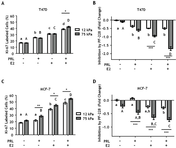
Figure 6. Stiff environments increase FAK-mediated hormone induced proliferation.

T47D and MCF-7 cells were plated on 12 or 75 kPa polyacrylamide gels coated with 200 μg/ml collagen-I in phenol-red free 5% charcoal stripped FBS for 24 h, serum starved for 24 h, and then treated with vehicle (DMSO 1:1000) or the FAK inhibitor, PF-573228 (1μM), for 1 h prior to ± PRL (4 nM), ± E2 (1nM) for 24 h. Cells were then stained with DAPI and Ki-67 antibody as described in Experimental Procedures. A, C. Effect of hormones on Ki67 staining, assessed by percentage of Ki-67 positive T47D (A) and MCF-7 (C) cells. B, D. Inhibition of proliferation by PF-573,228 compared to vehicle treated T47D cells (B) and MCF7 cells (D). Different letters represent significant differences within each stiffness (lower case, 12 kPa; upper case, 75 kPa). * represent significant differences between the same treatments at different stiffnesses: *p<0.05, **p<0.01, ***p<0.001.
