
Figure 5. Low-dose ICJ blocks TGF-beta induced EMT in breast cancer cells.
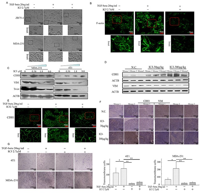
A. Morphological observation in ICJ treated breast cancer cells in the absence or presence of TGF-beta. After 48 hours of treatment, light-microscopic images were collected. Inset showed the details of the cellular morphological features. B. Confocal microscopic observation of the subcellular localization of F-actin in MDA-231 cells. The images showed the reduced polymerization and the re-organization of actin in response to ICJ treatment. C. Detection of EMT related markers in cells treated with ICJ (from 0.36 to 9.0μM) for 48 hours. D. Western blot verification of EMT-related markers by using the primary tumors (3 mice/group) obtained from the in vivo experiments mentioned in Figure 3. E. E-cadherin (CDH1) subcellular localization through Immuno-fluorescence analysis. The cells were treated with ICJ for 48 hours and CDH1 was visualized with FITC-labeled antibody. The nuclei were labeled by DAPI. F. Immunohistologicalchemical analysis of EMT molecular markers in primary tumors. The tissue samples were randomly selected from 3 mice in each group. G. Matrigel invasion assay of cells treated with ICJ alone or combined treated with ICJ and TGF-beta for 24 hours. The amounts of transmembrane cells were further quantified in five randomly selected microscopic fields.