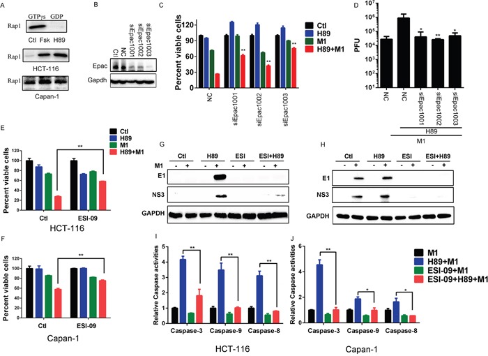
Figure 5. H89 activates Epac1 to increase oncolytic effects of M1.

A. Activated Rap1 determination. HCT-116 and Capan-1 cancer cells were treated with Forskolin (10μM) or H89 (10μM) for 24 hours and active Rap1 was detected with the Active Rap1 Detection Kit. GTPγs and GDP indicates positive control and negative control, respectively. B-D. The effects of Epac1. HCT-116 cancer cells were transfected with small interfering RNA (siRNAs) against Epac1. Epac1 expression levels were determined (B). Cell viabilities (72 hours post infection) and viral titers (48 hours post infection) were determined in the presence or absence of H89 after siRNA transfection (C and D). E and F. Cell viabilities determination with MTT assay after different treatment. HCT-116 and Capan-1 cancer cells were pretreated with ESI-09 or H89 or both for 1 hour and then infected with M1 (0.1 PFU/cell). Cell viabilities were determined 72 hours post infection. G and H. Determination of viral proteins E1 and NS3 with western blot. HCT-116 and Capan-1 cells were pretreated with ESI-09 or H89 or both for 1 hour, then M1 virus (1PFU/cell) was infected. Protein expression was determined 24 hours postinfection. I and J. Caspase-3, Caspase-9 and Caspase-8 activity assays (mean ± SD). HCT-116 and Capan-1 cells were plated on 96-well plates and M1 virus was infected for 72 hours in the presence or absence of H89 and/or ESI-09. *p<0.05; **p< 0.01. GAPDH, glyceraldehyde-3-phosphate dehydrogenase.
