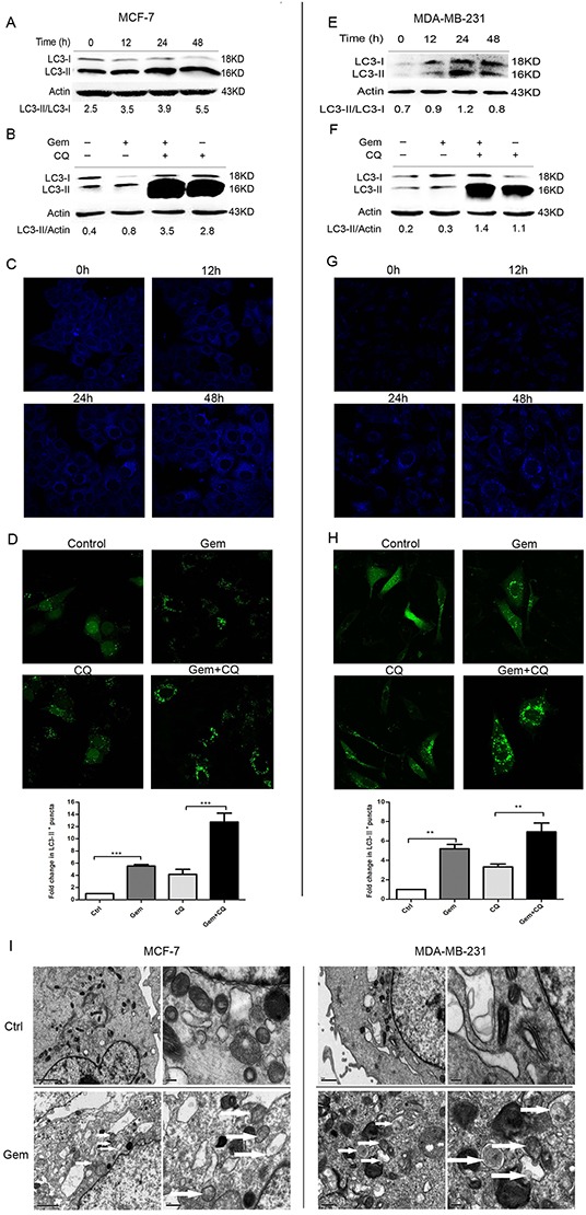
Figure 1. Gemcitabine induced autophagy both in ER-positive MCF-7 and ER-negative MDA-MB-231 cells.

A, E. The levels of LC3-I and LC3-II detected by Western blotting were quantified by densitometry using Image J software. The ratio of LC3-II to LC3-I was evaluated after treated with gemcitabine (MCF-7: 4 μg/ml; MDA-MB-231: 1 μg/ml) for 0 h (control), 12, 24 and 48 h. B, F. The ratio of LC3-II to actin was assayed after treated with gemcitabine, chloroquine (CQ) (MCF-7: 2.5 μM; MDA-MB-231: 5 μM), and gemcitabine plus CQ (added before gemcitabine treatment for 1 h) for indicated time (MCF-7: 48 h; MDA-MB-231: 24 h). C, G. Both the MCF-7 and MDA-MB-231 cells were exposed to gemcitabine for 0 h (control), 12, 24, 48 h, and treated with MDC (50 μmol/l) for 15 minutes, then fixed. The samples were analyzed by confocal microscope. D, H. Cells transiently transfected with GFP-LC3 plasmids were exposed to gemcitabine, CQ, gemcitabine plus CQ for 0 h (control) and 24 h. The confocal microscope was used to analyze GFP fluorescence. The number of LC3-II+ puncta was counted in at least 75 cells from random fields and the fold change (mean ± SE) was calculated by normalizing to the amount of control group. *, P<0.05;**, P<0.01;***, P<0.001. I. Observation of gemcitabine-induced autophagical vacuoles by transmission electron microscopy (TEM). MCF-7 and MDA-MB-231 cells were treated with gemcitabine for 0 h (control) and 24 h. Autophagical vacuoles with typical double-layer membrane containing remnants of organelles were highlighted by white arrows.
