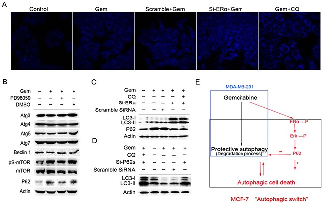
Figure 7. Excessive activation of the P62-mediated autophagic degradation by the phosphorylated ERα-ERK cascades may lead to the autophagic cell death induced by gemcitabine in ER positive MCF-7 cells.

A. MCF-7 cells that exposed to scramble siRNA or si-ERα for 48 h were treated by gemcitabine alone (4 μg/ml) or gemcitabine + CQ (2.5 μmol/L, added before gemcitabine treatment for 1 h) for another 48 h. Then the cells were treated with MDC (50 μmol/L) for 15 minutes and fixed. The samples were analyzed by confocal microscope. B. MCF-7 cells were treated by gemcitabine, gemcitabine+ PD98059 (30 μmol/L, added before gemcitabine treatment for 1 h) or DMSO for 48 h. Then total cell lysates were subjected to immunoblot analysis with indicated antibodies. The data represented a typical experiment conducted three times with similar results. C. MCF-7 cells were exposed to scramble siRNA or si-ERα for 48 h, and treated by gemcitabine or gemcitabine + CQ for another 48 h. The expression levels of LC3-I/II and P62 proteins were evaluated by Western blotting. β-actin was used as a loading control. D. MCF-7 cells were exposed to scramble siRNA or si-P62 for 48 h, and treated by gemcitabine or gemcitabine + CQ for another 48 h. The expression of LC3-I and LC3-II proteins was detected by immunoblot analysis and actin was used as a loading control. E. Working model for the “autophagic switch”. In ER negative MDA-MB-231, gemcitabine induced the protective autophagy. While in ER positive MCF-7 cells, the phosphorylation of ERα-ERK cascades induced by gemcitabine activated the P62-mediated autophagic degradation excessively, and as a result, it may lead to the collapse of cellular function and autophagic cell death. Inhibition of ERα-ERK cascades removed the excessive activation from the autophagic degradation process induced by gemcitabine, thus resulted in the autophagy that the degree was under a certain threshold, and was beneficial for the adaptation of cells in unfavorable conditions, thereby contributing to the cells survival.
