Figure 1.
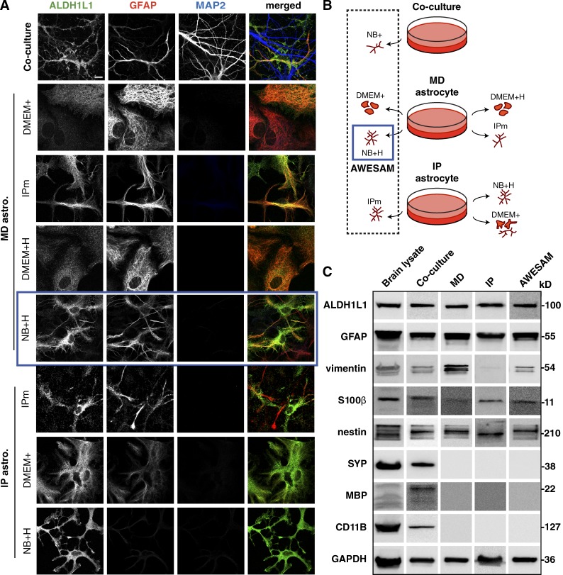
Different media yield morphologically different astrocytes. (A) Immunocytochemistry of DIV14 co-cultures and DIV16 astrocyte monocultures treated with different media. ALDH1L1 and GFAP label astrocytes; MAP2 labels neuronal dendrites. MD and IP astrocyte cultures were grown in different media as explained in B: Co-cultures were directly plated in NB+, whereas MD astrocyte monocultures growing in DMEM with 10% FCS (DMEM+) were shaken on DIV7 and then grown in DMEM+ for classical MD astrocytes, NB+ with HBEGF (NB+H), DMEM+ with HBEGF (DMEM+H), or the medium used in the Foo et al. (2011) IP astrocyte protocol (IPm), which is a 1:1 mix of DMEM and NB+H. IP astrocytes were generated as described by Foo (2013) and grown in IPm, NB+H, or DMEM+. In A and B, the blue square marks MD cultures grown in NB+H (AWESAM astrocytes), which we selected for further analysis because of the stellate morphology and ease of production. Images per experiment in A, from top to bottom: n = 6, 4, 8, 4, 8, 5, 4, and 6. Bar, 10 µm. (C) Immunoblots of adult rat brain lysate and DIV14 co-culture, MD, IP, or AWESAM astrocyte lysate. Antibodies targeted general markers of astrocytes (ALDH1L1 and GFAP), immature or reactive astrocytes (vimentin and GFAP), mature astrocytes (S100β), neurons (SYP), oligodendrocytes (MBP), and microglia (CD11B); GAPDH served as a loading control.

