Figure 8.
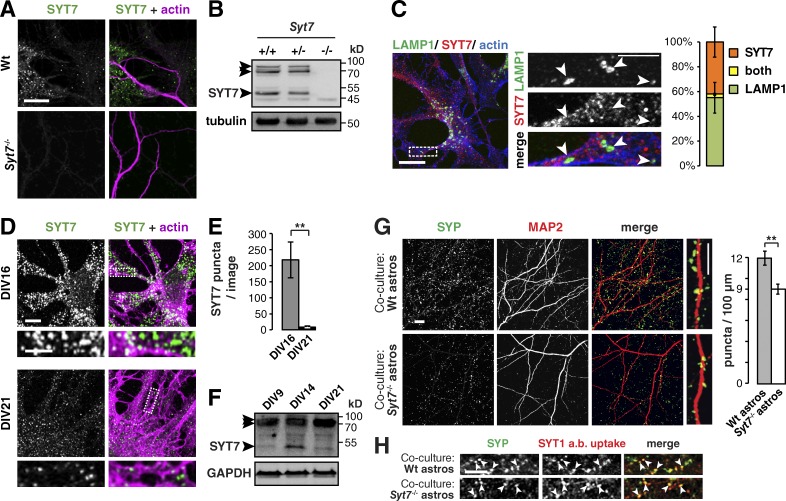
When astrocytes lack SYT7, neurons have fewer synapses. (A) Immunocytochemistry of DIV14 wild-type (Wt) and Syt7−/− co-cultures stained for actin and SYT7, where SYT7 puncta appear only in wild-type samples. Images per condition: n = 12. (B) Immunoblot showing three SYT7 splice isoforms at 47, 70, and ≈80 kD (arrowheads) in Syt7+/+ and Syt7+/− but not Syt7−/− adult brain lysate. Tubulin served as a loading control. (C) Immunocytochemistry and bar graph showing that the lysosomal protein LAMP1 (arrowheads) and SYT7 do not colocalize in DIV16 AWESAM astrocytes. The bar graph shows relative amounts of LAMP1 and SYT7 puncta and puncta positive for both (n = 6 images). (D) Immunocytochemistry comparing SYT7 expression levels in DIV16 and DIV21 AWESAM astrocytes (n = 5 images for DIV16, n = 3 images for DIV21). (C and D) Higher-magnification images of the boxed area (outlined by a dashed line) are shown in right (C) and bottom (D) panels. (E) Bar graph showing that astrocytes contain significantly more SYT7 puncta on DIV16 than DIV21. (F) Immunoblot showing that SYT7 expression is up-regulated in DIV14 AWESAM astrocytes versus DIV9 and DIV21. GAPDH served as a loading control. (G) Immunocytochemistry showing SYP puncta on dendrites (MAP2) of wild-type neurons grown on either wild-type astrocytes or Syt7−/− astrocytes. The bar graph shows that wild-type neurons have significantly fewer SYP puncta per 100-µm dendrite when grown on Syt7−/− astrocytes instead of wild-type astrocytes (n = 12 images for wild type and 14 images for Syt7−/−). (H) Antibody internalization assay showing that anti-SYT1 antibodies taken up during stimulation with 50 mM KCl solution colocalize with SYP (arrowheads) in DIV14 neurons grown on wild-type or Syt7−/− astrocytes (n = 6). Error bars indicate SEM, where **, P < 0.01 by unpaired two-tailed Student’s t test. Bars: (large view panels) 10 µm; (zoomed-in panels) 5 µm.

