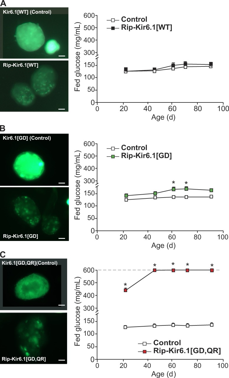
Figure 2.
Increased blood glucose in Kir6.1-GOF mice. (A–C, left) Representative intrinsic GFP fluorescence in freshly isolated islets from transgenic mice. High fluorescence in transgenic islets indicates the presence of the Kir6.1 transgene (Kir6.1[x], Control), and loss of fluorescence in transgenic β cells within islets after Cre-recombination (Rip-Kir6.1[x]) indicates actual expression of the transgene specifically in these cells. Bars, 10 µm. (right) Fed blood glucose over time after weaning in Rip-Kir6.1[WT] (A, black), single mutant Rip-Kir6.1[GD] (B, green), and double mutant Rip-Kir6.1[GD,QR] (C, red) with respect to their control littermates (white). Data represent mean ± SEM, n = 8–10 animals in each group. Significant differences (*, P < 0.05) between transgenic and control littermate mice are indicated (t test).

