
Figure 7.
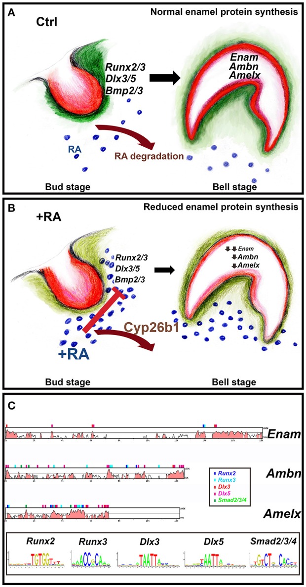
Model of how RA excess impairs enamel formation. Dental mesenchyme (green) and oral epithelium (red) have reciprocal interactions during tooth development (Balic and Thesleff, 2015) (A,B). (A) Normally the entire developing tooth is shielded from RA by actions of Cyp26 RA-degrading enzymes. This allows proper expression of Runx2/3, Dlx3/5, and Bmp2/3 in the mesenchyme condensing next to the oral epithelium at the bud stage (B). These targets are reduced by excess RA. Enam, Ambn, and Amelx exhibit strong reductions at the late bell stage, impairing enamel crystallization (C). The binding site motifs of Runx2/3, Dlx3/5, and Smad2/3/4 were obtained from the JASPAR database (http://jaspar.genereg.net) and used to find potential binding sites in conserved regions using the rVISTA database (http://genome.lbl.gov/vista/index.shtml).