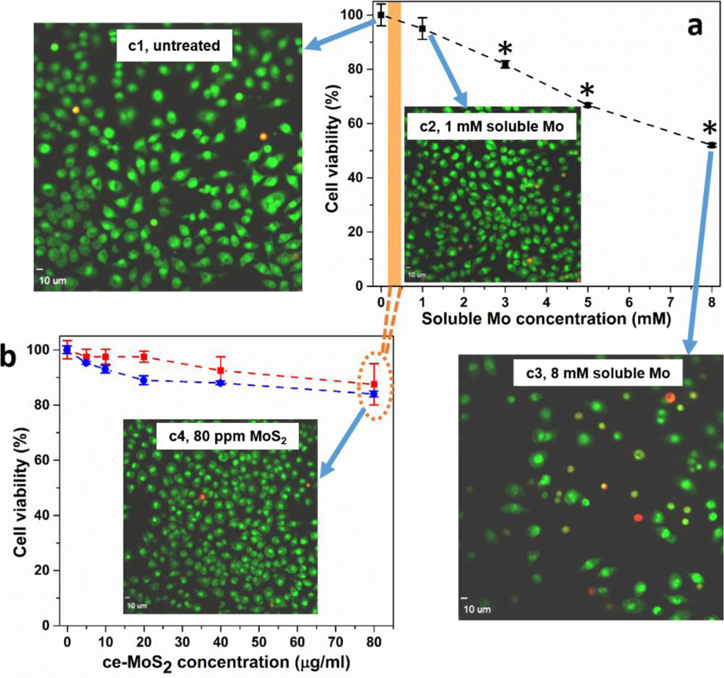
Figure 6.
Cytotoxicity for murine macrophages of ce-MoS2 nanosheets and soluble molybdate ions. Assessment of cell viability after exposure of murine macrophages to different concentrations of Mo salt (a) and ce-MoS2 sample (b). Following exposure to various amounts of Mo salt for 1 d or ce-MoS2 samples for 1 (red trace) or 2 days (blue trace), viability was assessed using dehydrogenase activity assay Wst 8. Concentrations above 3 mM Mo salt caused a significant decrease of viability (*p < 0.05). There was no significant cell death measured within 2 days of treatment in nanosheet samples. Images show visualization of cell death in murine macrophages using ethidium homodimer/Syto 10 stain after exposure to Mo salt or ce-MoS2 nanosheets for 24 hours. Macrophages were seeded into 96 well plates and imaged using an Olympus confocal microscope to visualize live (green) and dead (red) cells. Cells exposed to 8 mM Mo salt (c3) show significant loss in cell count and strong red fluorescence, while unexposed cells (c1), cells exposed to 1 mM Mo salt (c2), 80 µg/ml MoS2 nanosheets (c4) do not show toxicity, shown by strong green fluorescence. Images demonstrating uptake of ce-MoS2 by murine macrophages and human lung epithelial cells can be found in Supporting Information, Fig. S8

