Fig. 5.

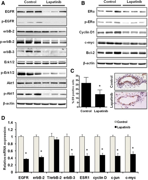
Lapatinib inhibits RTK and ER signaling pathways in vivo. a, b Total and phosphorylated protein levels of indicated markers in the mammary tissues (at 24 weeks of age) of mice from control and lapatinib (100 mg/kg/day for 8 weeks)-treated groups were detected using Western blotting. Protein samples from 3 mice in each group are shown. c Mammary tissue sections were prepared from 24-week-old control mice and mice treated with lapatinib (100 mg/kg/day) for 8 weeks. Percentages of ERα-positive cells were graphed as the means ± S.E. (* p < 0.05). Representative images of IHC analysis are shown with brown staining indicating ERα-positive cells. d mRNA levels of indicated markers in the mammary tissues of 24-week-old mice from control and lapatinib groups were quantified using real-time PCR. The relative mRNA expression of each indicated gene was graphed as means ± S.E. (* p < 0.05 versus the control for each gene)
