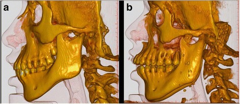
Fig. 4.

Conebeam CT scanning with 3D-reconstruction of craniofacial structures of two patients with Moebius. Patient no. 5 (a) and patient no. 4 (b). Both have a large maxillary prognathism in relation to the anterior cranial base (ACB) thus, having relatively retrognathic mandibles. In addition, the mandibular alveolar prognathism in relation to the mandibular base is large. Patient no. 4 (b) have severely proclined upper incisors with very divergent jaw-bases opening anteriorly and a marked reduction of the maxillary inclination in relation to the ACB and an anterior open bite
