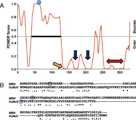
Fig. 2.

Disorder profile of GII.4 Sydney NS1-2 protein. a PONDR results showing predicted disordered region at the N-terminus of NS1-2 protein, the caspase cleavage site (yellow arrow; Cascleave), position of the H-box and NC motif (blue arrows), and the putative transmembrane domain (red arrow; PSIPRED) in the ordered region of the protein. The predicted phosphorylated threonine is indicated by a blue dot (DISPHOS). b Alignment of the disordered residues of MNV and HuNoV NS1-2 showing conserved motifs (Clustal Omega; * identical, : conserved substitutions, and . semi-conserved amino acids are indicated), and predicted phosphorylated threonine/tyrosine residues (blue circles; DISPHOS)
