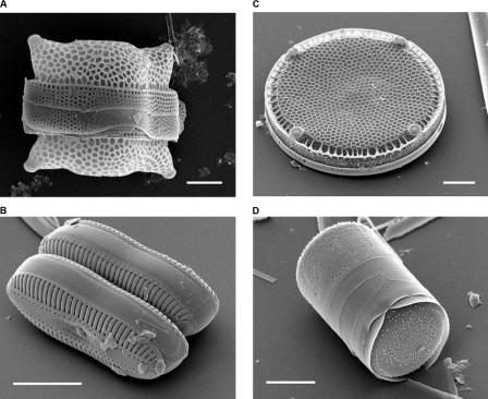
Figure 1. Scanning Electron Micrographs of Diatoms.
(A) Biddulphia reticulata. The whole shell or frustule of a centric diatom showing valves and girdle bands (size bar = 10 micrometres).
(B) Diploneis sp. This picture shows two whole pennate diatom frustules in which raphes or slits, valves, and girdle bands can be seen (size bar = 10 micrometres).
(C) Eupodiscus radiatus. View of a single valve of a centric diatom (size bar = 20 micrometres)
(D) Melosira varians. The frustule of a centric diatom, showing both valves and some girdle bands (size bar = 10 micrometres).
(Images courtesy of Mary Ann Tiffany, San Diego State University.)

