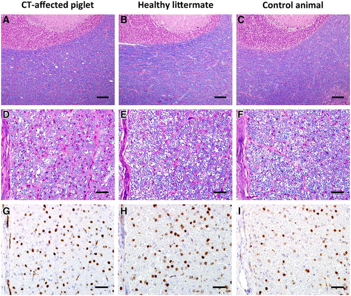
Figure 1.

Histological lesions in a CT-affected animal compared to a healthy littermate and a healthy control. Vacuoles in cerebellar white matter in affected animal (A), normal white matter in littermate (B) and control (C), LFB-HE, bar = 150 µm. Hypomyelination in white matter of the thoracic spinal cord in affected animal (D), normal myelination in littermate (E) and control (F), LFB-HE, bar = 40 µm. Detection of oligodendrocytes, increased staining intensity in affected animal (G), less intense staining in littermate (H) and control (I), Olig2-IHC, bar = 40 µm.
