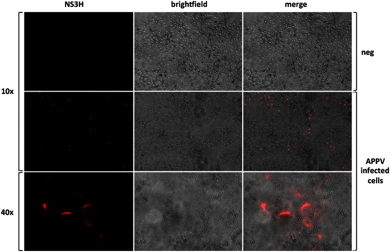
Figure 5.

Detection of APPV infected PK15 cells. A porcine anti APPV serum purified by NS3 affinity chromatography and a goat-anti swine Cy3 labelled polyclonal antibody was used for fluorescence staining. Cy3 fluorescence, brightfield and merge images are shown for uninfected and infected cells at 10× magnification. To resolve the perinuclear staining pattern, a cluster of positive cells is also shown at 40× magnification.
