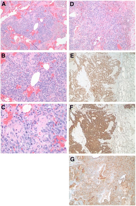
Fig. 2.

Images of the tumor from the lung. a-d Hematoxyalin and eosin-stained tumor sections from lung nodules. a Low power magnification (40X) of tumor demonstrated nodules of tumor cells can be seen in a background of abundant, fresh blood cells. b Medium power magnification (100X) showed the tumor cells are both epitheliod and spindle-shaped in appearance. The epitheliod morphology predominates in this area of the tumor. c High power magnification (400X) illustrated the tumor cells have prominent nucleoli. A mitotic figure can be seen in the center of the image confirming the tumor is mitotically active. d Another view of the tumor showing both epitheliod and spindle-shaped tumor cells, but in this section the spindle-shaped cells predominant (40X). e Tumor cells stained positive for CD34, a vascular marker (40X). f Tumor cells showed intense signal for CD31, another vascular marker (40X). g Factor VIII-related antigen, another endothelial marker commonly used to identify vascular tumors, demonstrated positivity in the tumor cells (40X)
