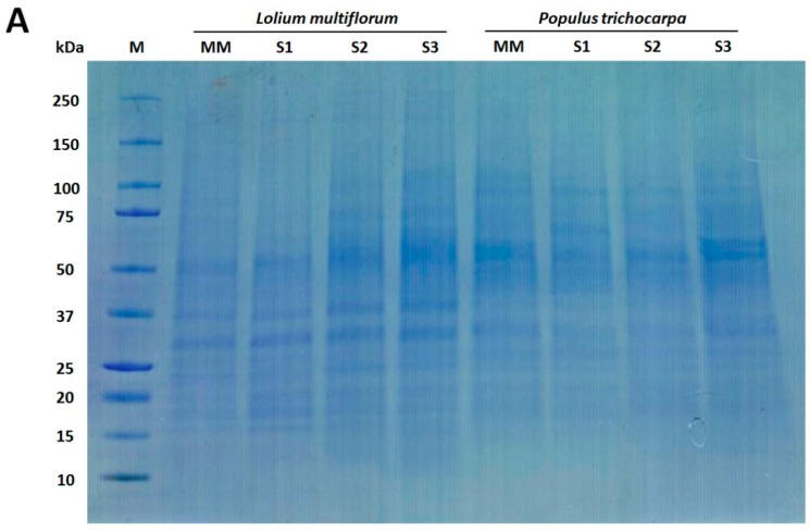
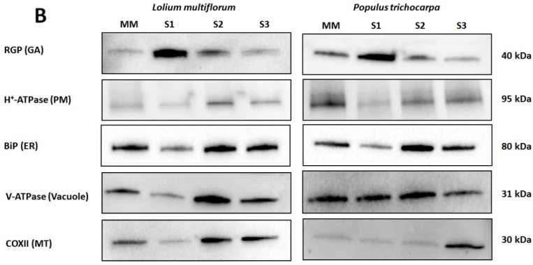
Figure 2.
Western blot analysis of Lolium multiflorum and Populus trichocarpa suspension cell culture (SCC) membrane fractions. Fractions (10 μg of total protein each) from MM, S1, S2, and S3 were separated by SDS-PAGE. (A) Coomassie blue staining of the total proteins separated on SDS-PAGE demonstrating the equal protein loading across the membrane fractions, (B) Western blots performed with anti-RGP (GA marker), anti-H+-ATPase (PM marker), anti-BiP (ER marker), anti-V-ATPase (vacuole marker), and anti-COX II (MT marker) antibodies. The molecular masses of the respective marker proteins are indicated on the right.


