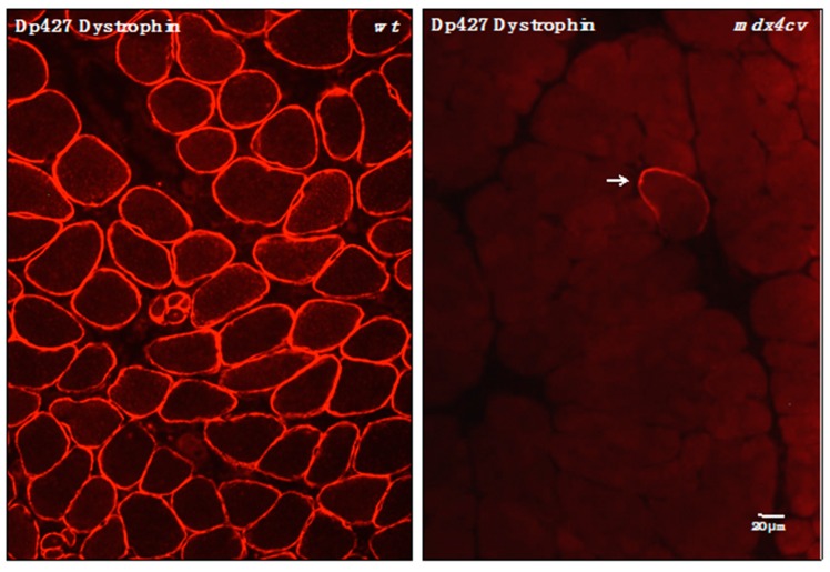
Figure 2.
Immunofluorescence microscopical localization of dystrophin isoform Dp427 in transverse cryosections from normal wild-type (wt) versus dystrophic mdx-4cv gastrocnemius muscle. Shown is the labeling of the sarcolemma in normal wt muscle using antibodies to dystrophin [71]. In stark contrast, the Dp427 isoform is almost completely absent from mdx-4cv muscle tissue. The arrow indicates a dystrophin-positive revertant fiber, which is extremely rare in the mdx-4cv mouse model of Duchenne muscular dystrophy [50].

