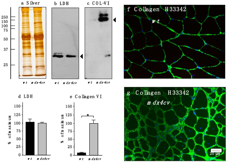
Figure 4.
Immunoblot analysis and immunofluorescence microscopical localization of collagen in normal versus dystrophic mdx-4cv gastrocnemius muscle. Shown is a representative silver-stained gel (a) and immunoblots (b,c). Lanes one and two represent total extracts from control wt muscle and dystrophic mdx-4cv skeletal muscle, respectively. Blots were labeled with antibodies to lactate dehydrogenase (LDH) (b) and collagen isoform COL-VI (c). Arrowheads mark the main immuno-labeled protein bands in individual panels. Graphical representations of the immuno-decoration levels for lactate dehydrogenase and collagen in normal versus mdx-4cv skeletal muscles are shown in panels (d,e): Student’s t-test, unpaired; n = 4; * p < 0.05. The immunofluorescence microscopy panels (f,g) show the labeling of the extracellular matrix in normal wt versus mdx gastrocnemius muscle, respectively, using antibodies to collagen COL-VI. In Dp427-deficient mdx-4cv muscle tissue the levels of collagen are greatly increased. Nuclei were stained with the DNA binding dye bis-benzimide Hoechst 33342 (H33342).

