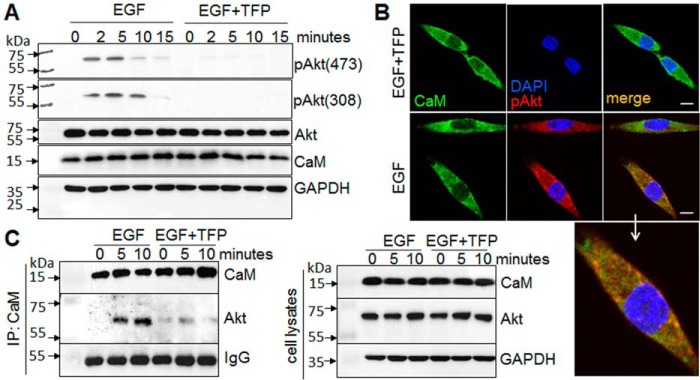
FIGURE 10.
CaM inhibition attenuates Akt activation. Pancreatic cancer cells, MiaPaCa-2, were exposed to serum-free media with or without TFP (10 μm) for 30 min and subsequently exposed to EGF (20 ng/ml) with or without TFP. A, Western blotting analysis of the phosphorylation of Akt at Ser-473 and Thr-308 in response to EGF for up to 15 min. B, confocal imaging analysis of EGF-induced Akt phosphorylation (pAkt, red) and co-localization of pAKT (red) with CaM (green) at 5 min. Enlarged image shows co-localization of pAkt with CaM. Scale bar, 10 μm. C, interaction of CaM and Akt, assessed by immunoprecipitation (IP) with CaM antibody followed by Western blotting analysis of CaM and Akt with specific antibodies. Representative results of at least three independent experiments are shown.

