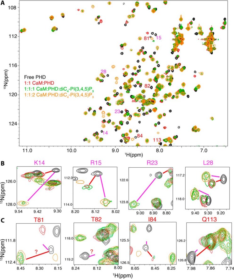
FIGURE 4.
A, overlay of 2D 1H-15N HSQC spectra obtained for a 15N-labeled Akt(PHD) sample (60 μm) in the free state (black) and when in complex with Ca2+/CaM (red). diC4-PI(3,4,5)P3 was then added at a 1:1:1 (green) and a 2:1:1 ratio (orange) (diC4-PI(3,4,5)P3/Akt(PHD)/CaM) followed by acquisition of 2D 1H-15N HSQC. Spectra of selected regions showing signals of residues affected by either binding of lipid (B) or CaM (B) are shown. Red and magenta lines indicate the positions of signals of CaM- or lipid-bound states, respectively. diC4-PI(3,4,5)P3 binding to Akt(PHD) displaces the C-terminal lobe of CaM as signals of residues in this region (β1/β2) shift to the lipid bound form (B). However, lipid binding is not able to displace the distant N-terminal lobe of CaM (C).

