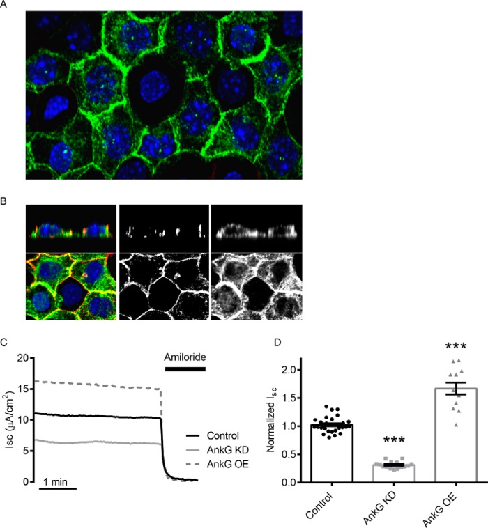
FIGURE 1.
AnkG expression in mCCD cells alters ENaC activity. A, confocal images of immunofluorescently labeled AnkG (green) in mCCD cells. B, magnified z stack images of mCCD cells with AnkG (green) and actin (red). The middle panel shows labeling of the cortical actin ring, and the right panel demonstrates AnkG at apical, lateral, and basal submembranous surfaces. C, representative short circuit current (Isc) recordings of ENaC activity with control (black), AnkG KD (light gray), and OE (dashed, dark gray). 10 μm amiloride is added at the end of the recording to determine the ENaC-specific contribution of the current recordings. D, summary of normalized Isc from several experiments. AnkG KD reduces ENaC current (0.31 ± 0.06 versus 1.00 ± 0.03, n ≥ 15, p < 0.001), and AnkG OE increases ENaC current (1.67 ± 0.11 versus 1.00 ± 0.06, n ≥ 12, p < 0.001).

