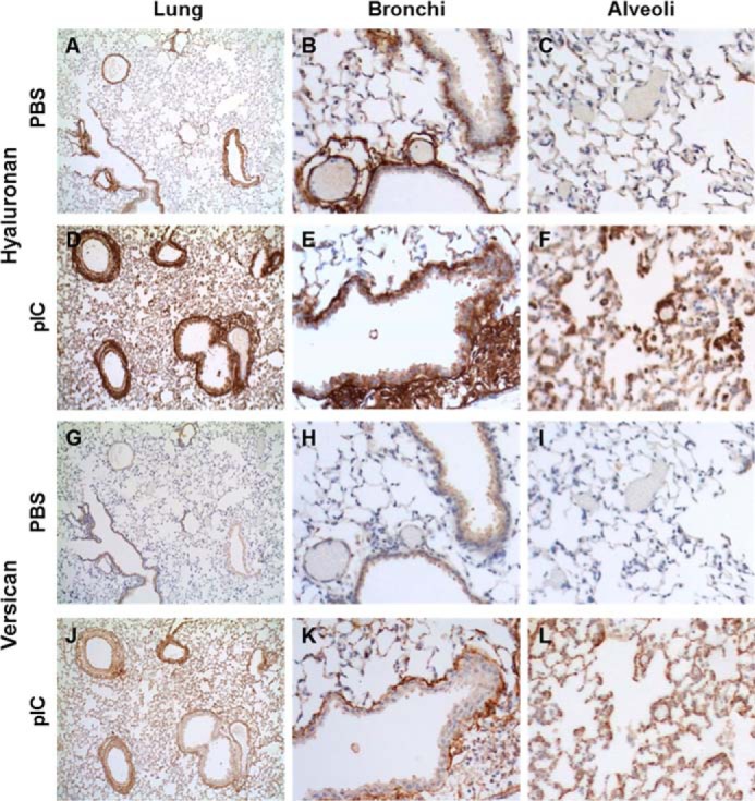
FIGURE 1.

HA and versican staining in PBS- (A–C and G–I) and poly(I:C)- (pIC) (D–F and J–L) instilled lungs. HA, which is present in areas surrounding bronchi and vasculature (B) and generally absent in alveolar spaces of unstimulated lungs (C), greatly increases in response to poly(I:C) stimulation (D), especially in areas enriched in infiltrating leukocytes (E), including the alveolar spaces (F). Versican, in contrast, is almost absent in PBS-treated lungs (G–I) but accumulates markedly in poly(I:C)-stimulated lungs (J), both in the peribronchial area (K) and in alveoli (L).
