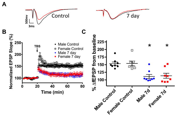
Fig. 1.
Ischemia impairs synaptic plasticity in juvenile mice. (A) Example fEPSP traces from control mice and mice 7 days after CA where red indicates pre-TBS trace and black indicates post-TBS trace. (B) Time course of fEPSP slope (mean±SEM) from control male mice (black circle), control female mice (white square), male (blue circle) and female (red square) 7 days after CA/CPR. Arrow indicates timing of theta-burst stimulation (40 pulses). (C) Quantification of change in fEPSP slope after 60 min following TBS normalized to baseline, set to 100%. *p<0.05 compared with control mice. (For interpretation of the references to color in this figure legend, the reader is referred to the web version of this article.)

