Figure 7.
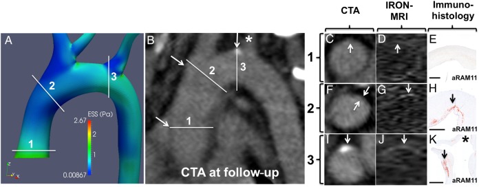
CFD, CTA, contrast MRI, and histopathological validation: (A) CFD of a 3D reconstructed aorta showing three convex curvature subsegments with high ESS (1) or low ESS (2 and 3) at baseline. (B) Multiplanar reformatting of the aorta by CTA at follow-up. Cross-sectional image of subsegments 1–3 by CTA (C, F, and I), IRON-MRI (D, G, and J), and histopathological examination (E, H, and K, aRAM11; bars represent 1000 µm) showing no plaque in subsegment 1 and inflamed plaque in subsegments 2 and 3. Of note, the asterisks in (B and K) correspond to the ostium of the left innominate artery.

