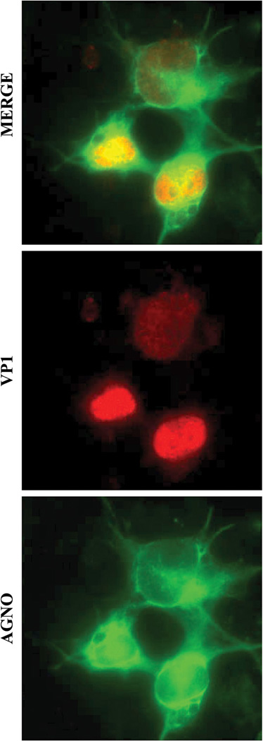
Fig. 1.
Cellular distribution of JCV agnoprotein in infected cells. Detection of agnoprotein and VP1 by immunocytochemistry was described previously (Sariyer et al., 2011). Briefly, SVG-A cells were transfected/infected with JCV Mad-1 strain and 15th day post-infection, cells were fixed with cold acetone on glass chambers and incubated with a primary anti-Agno polyclonal (Del Valle et al., 2002) and anti-VP1 (PAb597) monoclonal antibodies followed by incubation with a FITC-conjugated goat anti-rabbit and rhodamine-conjugated goat anti-mouse secondary antibodies and examined under a fluorescence microscope.

