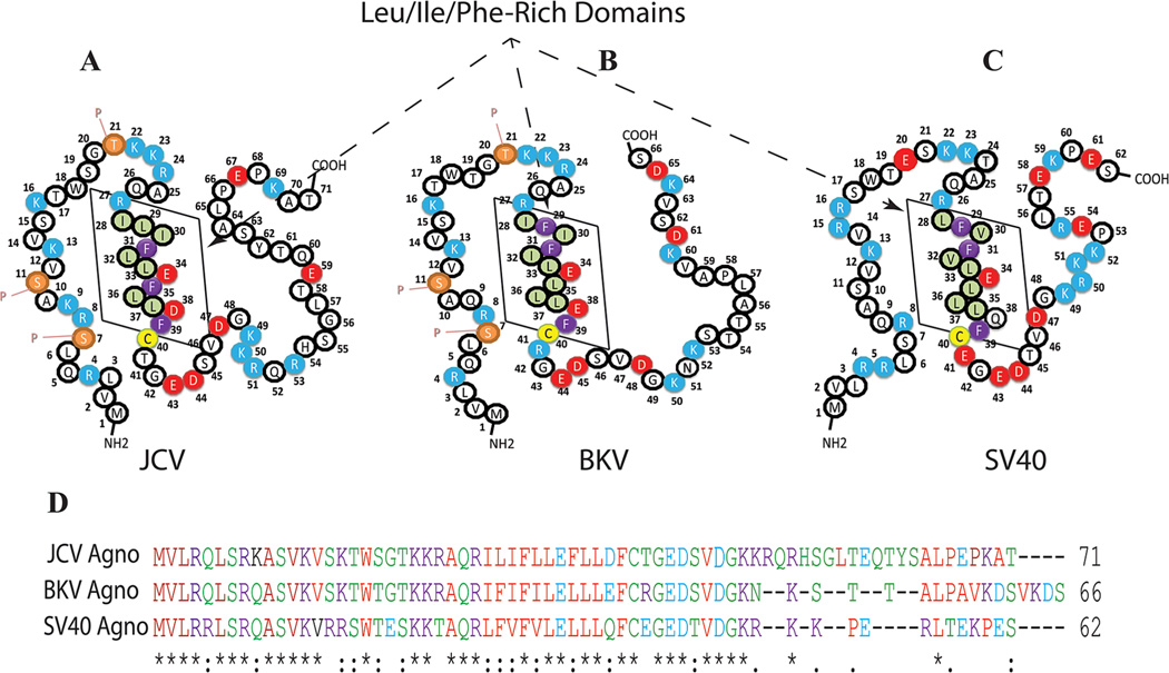
Fig. 3.
A–C: Primary structures of the JCV, BKV, and SV40 agnoproteins, respectively. The Leu/Ile/Phe-rich domain of each protein is indicated by a box. Phosphorylation sites in JCV and BKV agnoproteins are designated by the letter “P.” D: Amino acid sequence alignment of JCV, BKV, and SV40 agnoproteins.

