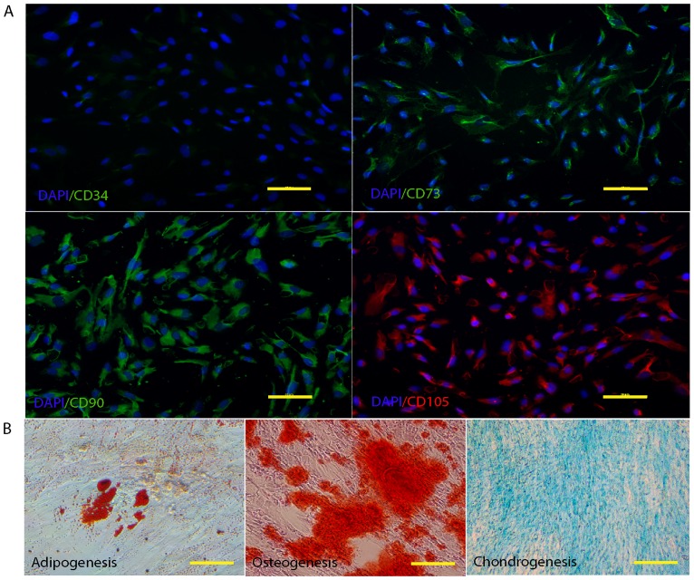
Fig 2. Characterization of hWJ-MSCs.
(A) Immunophenotype of MSCs, immunofluorescent micrographs staining expression of MSC markers (CD73, 90, and 105), Nuclei were counterstains with DAPI (blue). Cells were negative for hematopoietic marker (CD34). Scale bar = 20 μm. (B) Differentiation of hWJ-MSCs to mesodermal linage cells. The cells were induced to undergo adipogenic, osteogenic, and condrogenic differentiation.

