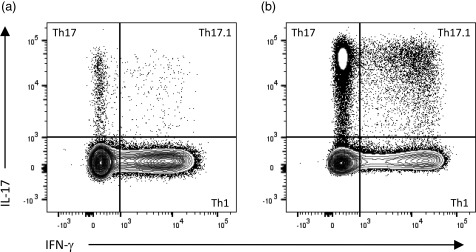
Figure 1.

Intracellular cytokine staining to identify T helper type 1 (Th1), Th17 and Th17.1 cells. In comparison to a healthy control sample (a), the proportion of Th17 and Th17.1 cells can be increased substantially in samples from patients with clinically isolated syndrome (CIS), particularly in the setting of active disease (b). Plots represent CD3+CD4+ T cells gated on live, single‐cell peripheral blood mononuclear cells (PBMC), following a 4‐h stimulation with phorbol myristate acetate (PMA)/ionomycin in the presence of brefeldin A (BD leucocyte activation cocktail with GolgiPlug).
