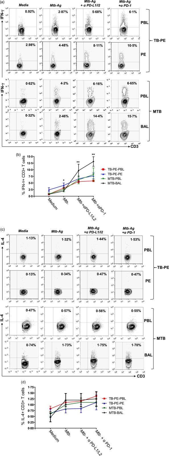
Figure 3.

Mycobacterium tuberculosis stimulation combined with programmed death‐1 (PD‐1)–PD‐ligand (PD‐L2) blockade efficiently increases the frequency of T helper type 1 (Th‐1) cells, and sparing Th‐2 T cells. Mononuclear cells from peripheral blood (PB) and pleural fluids of tuberculosis‐pleural effusion (TB‐PE) patients (n = 4); PB and bronchoalveolar lavage of miliary tuberculosis (MTB) patients (n = 4) were blocked with α‐PD‐1 alone or α‐PD‐L1 and α‐PD‐L2 in combination with purified monoclonal antibodies (mAbs) blocking antibodies for 72 h with or without M. tuberculosis antigen (WCL = whole cell lysate). T cells from cultured cells were analysed for M. tuberculosis‐specific cytokine production by flow cytometry. Lymphocytes were gated based on their scatter profile and gated further on the basis of CD3 expression. Representative fluorescence activated cell sorter (FACS) plots show production of (a) interferon (IFN)‐γ and (c) interleukin (IL)−4 among study groups. Each point represents the mean percentage ± standard error of the mean from each group for (b) IFN‐γ and (d) IL‐4 production in various culture conditions. *P = 0·01; **P = 0·001. [Colour figure can be viewed at wileyonlinelibrary.com]
